

Organize your questions and next steps, then save your worksheet.
Summary and next steps
Cataracts, glaucoma, AMD and diabetic retinal disease involve different care questions. Use the guide to prepare a focused conversation rather than decide that vision is safe.
- List the diagnoses or symptoms you need explained.
- Bring your medicine list and any previous eye treatment.
- Ask for the next examination, treatment and support steps.
Editorial maintenance: October 24, 2026. Summary layout, related links and technical metadata updated. This is not a full clinical review.
You know you’ve crossed a line when “reading the fine print” means holding the menu at arm’s length, then squinting, then asking your dinner companion to read it out loud like it’s a group project. If you’re over 60 and starting to feel like your eyes are staging a quiet rebellion, trust me—you’re in good company.
If you cannot read a medicine label clearly, do not guess the drug name or dose. Ask a pharmacist or caregiver to read it with you and request an accessible label or another reliable format. New difficulty reading also deserves an eye assessment.
Treatment and vision rehabilitation can help many people protect function and independence. The outcome depends on the cause and stage of disease; neither this guide nor a symptom checklist can determine that your vision is safe for driving.
This guide is your no-jargon, no-eye-rolls cheat sheet to the four big age-related eye diseases. We’re talking real talk: what warning signs to watch for, what’s normal aging versus “hey, you should call someone,” and how much this stuff actually costs (spoiler: Medicare may have your back more than you think).
You’ll also find some surprisingly simple daily habits that can actually protect your sight—not in a “someday you’ll thank me” way, but in a “start this week and it makes a difference” kind of way.
This guide explains common conditions and questions for an eye appointment. Use the urgency reminders below to seek care promptly, not to rule out an emergency or postpone assessment.
Your eyes have served you well—let’s return the favor.
Important: This article is for education, not diagnosis. If you notice sudden vision changes, eye pain, or a “curtain” over your sight, treat that as an emergency and contact an eye doctor or emergency services right away.
Why Your Vision Changes After 60
After 60, your eyes are doing exactly what the rest of your body is doing: aging, adapting, and occasionally complaining. The lens inside your eye slowly stiffens and yellows, fluid drainage pathways can clog, the macula (your fine-detail center) can thin, and tiny blood vessels may leak if you live with diabetes. None of this means automatic blindness—but it does mean you can’t rely on “I see fine” as proof that everything is okay.
Age-related eye disease can be present before you notice a change. Early assessment can identify treatable causes and establish monitoring when damage cannot be reversed. Symptoms alone do not distinguish normal aging from disease.
For example, gradually worsening night glare can be caused by cataracts and may make driving unsafe. Discuss the change with an eye professional rather than assuming that a new glasses prescription will solve it. This is an illustrative scenario, not a reported patient outcome.
Plan regular comprehensive eye exams. The National Eye Institute recommends a dilated exam every 1–2 years for adults over 60; diabetes, high blood pressure, diagnosed eye disease, or other risks may require more frequent review. Ask your eye doctor for your individual interval.
- Seek care immediately if you have sudden vision loss, a dark curtain, new flashes of light, or severe eye pain.
- Contact an eye doctor right away for new wavy lines or a new central gray spot. A sudden increase in floaters, flashes, or a curtain needs immediate assessment; do not wait 1–2 weeks.
- If you have no new symptoms, check your last exam date and your eye doctor’s recommended interval. Arrange a routine appointment if you are due; new symptoms need their own prompt assessment.
Neutral action: Screenshot this list or write it down, and ask your eye clinic where your symptoms fit before you schedule.
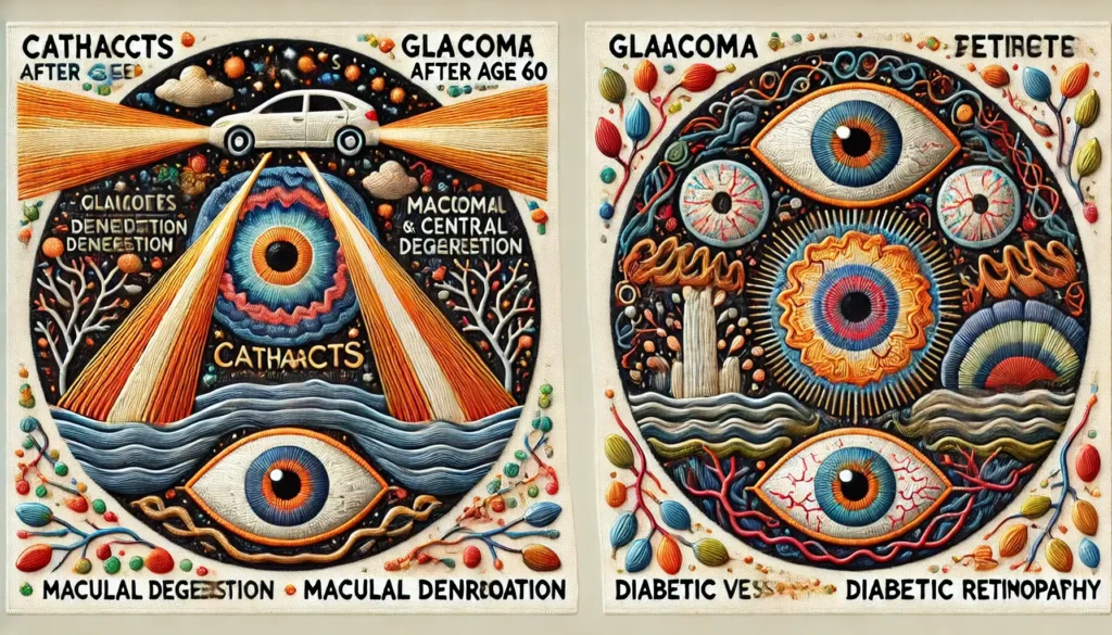
Infographic: Your Four Main Eye Diseases After 60
Cataracts
Main issue: Cloudy lens.
Typical age: 60+.
Red-flag: Glare and halos make night driving unsafe.
Glaucoma
Main issue: Optic nerve damage; pressure may be high or normal.
Typical age: 60+ risk jumps.
Red-flag: Often none—needs comprehensive eye exams, not pressure checks alone.
Macular Degeneration
Main issue: Central retina thinning.
Typical age: 65+.
Red-flag: Straight lines look wavy on the page.
Diabetic Retinopathy
Main issue: Leaking retinal blood vessels.
Typical age: Any age with diabetes; risk climbs after 60.
Red-flag: Blurry or patchy spots in both eyes.
Use this as a mental “map.” The rest of the article simply walks you through each box in more detail.
Cataracts: Cloudy Vision, But Very Treatable
Cataracts are why car headlights feel like searchlights and why your once-crisp newspaper now looks like it’s been printed through a fog. A cataract is just a cloudy natural lens. It usually grows slowly, which is why people often blame “dirty glasses” or “old light bulbs” instead.
Cataracts become more common with age. Surgery is the treatment that removes a cataract, but the decision depends on its effect on daily activities and the likely benefits and risks for your eyes.
Cataract surgery can improve vision and daily activities when cataract is the main cause. Retinal, optic-nerve, and other conditions affect the likely benefit; ask your surgeon what improvement is realistic for you.
Common cataract symptoms after 60
- Hazy, filmy, or “dirty” vision even with new glasses.
- Glare and halos from headlights or bright lights.
- Colors look dull or yellowed.
- Frequent prescription changes without real improvement.
Cataracts do not usually cause pain or redness. If you have pain, that’s a separate, urgent problem and needs immediate care.
Cataract surgery, cost, and coverage questions (US, 2026)
Cataract surgery removes the cloudy natural lens and usually replaces it with an intraocular lens (IOL). It is commonly an outpatient procedure; appointment length and recovery vary. Ask for an itemized quote covering the surgeon, facility, anesthesia, lens, testing, and follow-up rather than relying on an unsourced national cash-price range.
For covered outpatient cataract surgery under Original Medicare, the Part B deductible and usually 20% of approved charges apply. Ask which lens features and optional refractive services would be noncovered and separately priced. Medicare Advantage and supplemental coverage can change your share. See Medicare’s cataract surgery coverage guidance.
- Watch for glare, halos, and faded colors.
- Ask your surgeon to show you standard vs premium lens options and costs.
- Confirm how much Medicare or your insurer pays before booking a surgery date.
Apply in 60 seconds: Write down one activity cataracts are interfering with (e.g., night driving), and bring that sentence to your next eye appointment.
Show me the nerdy details
Inside your eye, the natural lens is made mostly of water and proteins arranged in a very precise pattern. With age, oxidative stress and UV exposure change these proteins so they clump and scatter light. Different patterns of clouding (nuclear, cortical, posterior subcapsular) cause different symptoms: nuclear cataracts often change your glasses prescription first, while posterior subcapsular cataracts create intense glare. The artificial intraocular lenses used in surgery are usually acrylic or silicone and can be monofocal (single distance), multifocal, toric (for astigmatism), or light-adjustable. These choices drive both your visual outcome and your out-of-pocket costs.
Glaucoma: The “Silent Thief of Sight”
Glaucoma is a group of diseases where damage to the optic nerve slowly steals your side vision. You don’t feel it happening. You don’t usually see it happening. That’s why many people discover it late—during a routine pressure check they almost skipped.
Glaucoma risk increases with age, family history, and certain other factors. High eye pressure is a risk factor, but glaucoma can also occur at normal pressure. A comprehensive exam assesses the optic nerve and, when needed, visual fields; pressure testing alone cannot rule it out. See NEI guidance on glaucoma and eye pressure.
Someone seeking new reading glasses may be unaware of glaucoma-related damage. Regular assessment matters because the usual open-angle form often has no early symptoms; treatment aims to limit further damage rather than restore vision already lost.
Key risk factors you can’t ignore
- Age over 60.
- Family history of glaucoma, especially parents or siblings.
- African, Hispanic/Latino, or Asian ethnicity.
- Thin corneas, high eye pressure, or very high/low eye prescriptions.
- Long-term steroid use (including some inhalers and creams).
Glaucoma care often means lifelong monitoring, pressure-lowering drops, and sometimes laser or surgery. Think of it as managing blood pressure for your optic nerve.
- If you’re over 60, ask explicitly, “Did you check me for glaucoma today?”
- Write down your target eye pressure if your doctor sets one.
- Set a phone reminder to use glaucoma drops at the same time every day.
Apply in 60 seconds: Add “Ask about my optic nerve and pressure” as a note in your calendar for your next eye visit.
Macular Degeneration: Slow Loss of Central Vision
Age-related macular degeneration (AMD) damages the macula, the tiny central area of your retina responsible for sharp vision. You can think of it as a worn patch on the sensor of a camera—edges might look okay, but faces, print, and road signs become blurry or distorted.
AMD is a leading cause of vision loss in older adults. Early disease can be present before symptoms; new central distortion warrants prompt assessment.
A new missing patch in print or new wavy lines can signal a macular problem. Contact an eye doctor right away rather than assuming a glasses change. Only an examination can determine the cause and stage.
Dry vs wet AMD
- Dry AMD: More common; gradual thinning and “waste” deposits (drusen) under the macula. Vision changes are often slow.
- Wet AMD: Less common but more aggressive; abnormal blood vessels leak under the macula, causing rapid distortion and central blind spots.
Treatment depends on AMD type and stage. Wet AMD commonly requires anti-VEGF injections on an individualized schedule. Ask whether stage-specific supplements or treatment for geographic atrophy are relevant, rather than taking generic eye vitamins or assuming all dry AMD is untreatable. The FDA describes an approved geographic-atrophy treatment in its IZERVAY review. Discuss expected benefit and injection risks with a retina specialist.
- If your clinician recommends Amsler monitoring, use a proper grid as instructed, often daily for AMD, and report new distortion immediately. See AAO guidance on AMD monitoring.
- Talk to your doctor before starting any “eye vitamins” or supplements.
- If you already get injections, don’t skip visits—even when vision seems stable.
Apply in 60 seconds: Ask your eye clinic for an Amsler grid and instructions. Check each eye separately using your usual reading correction and the recommended distance; do not wait for the next check to report a new change.
Diabetic Retinopathy: When Blood Sugar Hurts the Eyes
If you live with diabetes, your eyes are quietly keeping score. High blood sugar over time can weaken and leak the tiny blood vessels in your retina, leading to diabetic retinopathy (DR). This can cause blurring, dark patches, or even sudden vision loss if bleeding or swelling is severe.
CDC modeled estimates put diabetic retinopathy at approximately 9.6 million U.S. residents across all ages in 2021, including 1.84 million with vision-threatening disease. These are historical population estimates, not a personal risk calculation. Risk varies with diabetes duration and other clinical factors.
Diabetic retinopathy may progress without noticeable symptoms. Keep the retinal screening and treatment schedule agreed with your diabetes and eye-care teams, even when reading vision seems good.
What you can do if you have diabetes
- Keep your A1C, blood pressure, and cholesterol as close to target as safely possible.
- Schedule yearly dilated retinal exams; sooner if your doctor recommends.
- Report any new floaters, patchy shadows, or sudden blur immediately.
- Ask whether you need retinal photographs or OCT scans at baseline.
- Pair your annual diabetes review with an annual eye check in your calendar.
- Ask for copies of your eye images to track progress over time.
- Talk with your eye specialist before any major diabetes medication change.
Apply in 60 seconds: If you have diabetes, open your calendar app and create a recurring “eye and A1C week” reminder once a year.
Cost, Coverage, and Medicare Questions for Eye Disease Treatment (US, 2026)
Even if you’re emotionally ready for treatment, the financial side can feel like another diagnosis. Cataract surgery, glaucoma lasers, macular injections, and diabetic eye treatments all involve different billing codes, prior authorization rules, and coverage tiers.
For 2026, the Original Medicare Part B annual deductible is $283. For covered outpatient cataract care, patient cost sharing is based on approved charges after the applicable deductible, not a retail price. Facility and professional charges may be separate, and noncovered refractive upgrades need their own quote.
For retinal injections, confirm the drug and procedure codes, medical-benefit coverage, any plan authorization, the clinic’s network status, and your expected share per visit. An uninsured drug charge, an insured copay, and the complete treatment-visit price are different figures. Ask about assistance before delaying medically recommended care.
| Service | Quote to request | Common coverage notes |
|---|---|---|
| Standard cataract surgery + basic lens (per eye) | Complete itemized per-eye quote | Often covered by Medicare/insurance after deductible, at ~80% of approved amount. |
| Premium or multifocal lens upgrade | Incremental noncovered upgrade price | Frequently not covered; paid out-of-pocket. |
| Anti-VEGF injection (wet AMD/DR) | Drug and administration charges separately | Plan-dependent; subject to prior authorization and copays. |
| Glaucoma laser or surgery | Exact procedure and facility quote | Often covered as medically necessary; out-of-pocket depends on deductible and coinsurance. |
This is a quote checklist, not a current national price survey. Confirm the exact procedure, drug, service setting, covered charges, and noncovered extras.
Neutral action: Save this table and confirm each line item with your insurer and clinic’s billing team before scheduling any procedure.
- Ask, “Is this covered under my medical plan or my vision plan?”
- Confirm your Part B deductible and coinsurance before cataract surgery.
- For injections, ask which drug and billing code will be used.
Apply in 60 seconds: Grab your insurance card and write down your deductible and typical coinsurance percentage on a sticky note near your phone.
Daily Habits That Protect Your Vision After 60
Here’s the hopeful part: small daily choices genuinely influence how quickly age-related eye diseases progress. You can’t change your birthdate or your genes, but you can change what you eat, how you move, and how you manage chronic conditions.
A varied diet can support general health alongside smoking cessation and management of blood pressure and diabetes. It does not replace prescribed eye treatment, and this guide does not promise a percentage reduction in AMD risk from a particular diet.
- Quit smoking and ask your care team about cessation support. Cutting down is a step toward quitting, not equivalent protection.
- Wear UV-protective sunglasses whenever you’re outdoors, even on cloudy days.
- Control blood pressure, cholesterol, and blood sugar with your primary-care team.
- Move your body most days of the week; even 20–30 minutes of walking helps circulation.
- Use task lighting and high-contrast materials at home to reduce eye strain.
For example, planning meals around vegetables, beans, fish where appropriate, and other foods recommended by your care team may make healthy eating easier. This is a practical suggestion, not a report of measured patient outcomes.
- Pair eye-friendly foods with something you already enjoy.
- Keep one pair of good sunglasses near your door, another in the car.
- Measure your home lighting once and upgrade the darkest spots.
Apply in 60 seconds: Add “greens + something orange” to your next grocery list as a visual-health reminder.
How to Prepare for Your Next Eye Appointment
Going to an eye doctor can feel like speed dating: a lot of questions, a short window, and you walk out thinking of three things you forgot to say. A little prep work turns that rushed visit into a real strategy session for your vision.
If you’re in the United States, it’s worth knowing that many routine eye exams fall into a gray zone: some are billed as “medical” (e.g., monitoring glaucoma, AMD, DR) and go through Medicare Part B or private insurance; others are “vision” exams (mostly refractive) and use a separate vision plan. That’s why you might see different copays and deductibles for what feels like the same visit.
Before you compare cataract surgery centers or retina clinics, gather:
- Your insurance card(s) and a photo of the back with customer-service numbers.
- A list of all eye medications and drops, plus any blood thinners or diabetes meds.
- Past eye surgery dates, if any (LASIK, cataract, retinal procedures).
- Your priorities: night driving, reading, computer work, or hobbies like sewing.
- Recent lab results (A1C, blood pressure summary) if you have chronic conditions.
Neutral action: Keep this list in a folder or note on your phone and bring it whenever you ask for a quote or second opinion.
In the UK or other countries with national health systems, your pathway may run through your general practitioner, who refers you to hospital eye services. Knowing your system’s referral rules—and waiting times—helps you decide if you need private care for faster access or if the standard path is enough.
- Write down your top three vision frustrations.
- Bring all current glasses and contact lens boxes.
- Ask, “If this were your eyes, what would you do in the next year?”
Apply in 60 seconds: Start a small “eye file” (physical or digital) and drop in your last exam summary, so you’re not scrambling before the next one.
Emergency Eye Symptoms You Should Never Ignore
Most age-related eye changes are slow and polite. Some are not. These are the situations where you should treat your eyes the way you’d treat chest pain: not tomorrow, now.
- Sudden vision loss in one or both eyes.
- A dark curtain or shadow moving across your vision (possible retinal detachment).
- New, intense flashes of light or a sudden “shower” of floaters.
- Severe eye pain, redness, halos around lights, and nausea (possible acute glaucoma attack).
- Sudden double vision or trouble moving one eye.
If any of these appear, bypass routine booking systems. Use your eye doctor’s emergency number, an emergency eye service or the emergency department—whatever gets you seen fastest and safely. Your future vision may depend on hours, not days.
Do not wait for a weekday appointment if a curtain, sudden shadow, flashes, or a sudden increase in floaters appears. The NEI advises seeing an eye doctor or emergency department right away for possible retinal detachment.
Living Well With Low Vision and New Technologies
Even with the best medicine, some people over 60 end up with permanent vision loss. That doesn’t mean life is over. It does mean you may need new tools, new habits, and sometimes new technology.
Low-vision specialists can help you use magnifiers, telescopic glasses, large-print devices, high-contrast settings on phones and tablets, and clever lighting setups at home. A few clever changes—like bump-on stickers on appliances and bold markers for medication labels—can cut daily frustration dramatically.
If you are interested in investigational implants or other new technologies, ask your retina specialist about the evidence, eligibility, local availability, regulatory status, and training involved. Research results should not be presented as a promise of restored reading vision for every patient.
Vision rehabilitation can teach strategies for making better use of remaining vision. Ask for an assessment rather than assuming that larger glasses alone are the only option.
- Ask your eye doctor for a “low-vision rehab” referral if print feels too hard.
- Experiment with phone accessibility settings: larger text, bold fonts, voice control.
- Consider support groups—vision loss is emotional as well as physical.
Apply in 60 seconds: On your phone, open accessibility settings and bump your text size one notch higher as a test.
Short Story: Finding Hope in a Waiting Room
Fictional illustration: Ellen, 74, returns to the retina clinic after a year of treatment for wet AMD. She is anxious about a new change in her better eye. A man across from her asks what to expect at his first visit.
“I’m scared I won’t be able to drive my kids,” he admitted. Without thinking, she said, “I’ve been getting injections for a year. I still read, boss my granddaughter, and terrorize my book club. You’ll learn the drill.” They both laughed, a little too loudly. Later, as she left, she realized something had shifted. Her eyes weren’t “failing” in isolation; she was part of a room full of people quietly fighting for their vision, one appointment at a time. The fear didn’t vanish, but it became something she could walk into instead of run from.
FAQ
Q1. How often should I get my eyes checked after 60?
The NEI recommends a comprehensive dilated eye exam every 1–2 years for adults over 60. Diabetes, high blood pressure, diagnosed eye disease, or other risks can require more frequent care; use your eye doctor’s individual schedule. New symptoms should not wait for a routine exam.
Next step: Check your last exam date and ask the clinic when you are due.
Q2. Does Medicare cover routine eye exams for seniors?
Original Medicare (Part A and B) generally doesn’t cover routine “check-your-glasses” eye exams, but it does cover medically necessary visits—for example, monitoring cataracts significant enough to affect safety, glaucoma, AMD, or diabetic eye disease. Many Medicare Advantage plans and separate vision plans offer additional routine coverage.
60-second action: Call the number on your insurance card and ask, “What eye exams are covered for me this year, and what is my copay?”
Q3. How do I know if my cataracts are “bad enough” for surgery?
There’s no single number on a chart that flips the switch. Instead, doctors look at how cataracts affect your daily life: night driving, reading, recognizing faces, work, or hobbies. If glasses no longer solve your problems and your cataracts are the main cause, surgery becomes a reasonable option.
60-second action: Write down three tasks that your vision makes difficult now; bring that list to your next eye visit and ask your doctor to comment on each one.
Q4. I have diabetes but good vision. Do I really need annual retinal exams?
Yes. Diabetic retinopathy can be quite advanced before you notice any blur. Regular retinal exams allow your doctor to catch small leaks, swelling, or new vessels early, when treatment is easier and more effective.
60-second action: Pair your retinal exam with another yearly event—an anniversary, tax season, or birthday—so you remember to schedule it consistently.
Q5. Can lifestyle changes really make a difference if I already have AMD or glaucoma?
They won’t replace medical treatment, but they can support it. Not smoking, eating an eye-friendly diet, controlling blood pressure and blood sugar, and using UV protection can reduce additional damage and help treatments work as well as possible.
60-second action: Choose one habit—such as wearing sunglasses whenever you step outside—and commit to improving that for the next two weeks.
Q6. What can I do if I can’t afford the copays for injections or surgery?
Many clinics have payment plans, social workers, or access to drug assistance programs from manufacturers. Some charitable organizations help with costs for eligible patients. Talk to the billing office before you cancel or delay treatment; they would rather help you find a safe solution than see your vision deteriorate.
60-second action: Before your next visit, call the clinic and say, “I’m worried about affording my treatment. Is there someone I can speak with about payment options or assistance programs?”
Optional video: Open the video on YouTube (external site).
Optional video: Open the video on YouTube (external site).
Summary and Next Steps
Let’s close the loop on that first question: “Are my aging eyes just annoying—or truly at risk?” The honest answer is usually “both.” Cataracts, glaucoma, macular degeneration, and diabetic retinopathy become more common after 60, but you’re not a passenger. With regular exams, smart questions, realistic cost planning, and a few surprisingly simple habits, you can tilt the odds strongly in favor of keeping the vision that lets you read, drive, cook, and recognize the faces you love.
In the next 15 minutes, you can:
- Book (or at least calendar) your next dilated eye exam.
- Read the urgent warning signs and seek care without completing a checklist first.
- Start an “eye file” with your last exam note, insurance info, and a list of questions.
Your eyes have carried you through millions of pages, roads, and faces. They’re worth a plan, not just a wish. Start with one small action today, and let your future self—still reading, still recognizing, still moving through the world with confidence—say thank you.
Last updated: October 15, 2026. Medical urgency, examination intervals, glaucoma screening, AMD monitoring, and U.S. coverage information were checked against linked official sources. This educational review does not establish an individual diagnosis or treatment plan.
🔗 Early Warning Signs of Cataracts in Your 60s and 70s (When to See an Eye Doctor): 7 Scary Red Flags I Nearly IgnoredPUT THIS GUIDE TO WORK · INTERACTIVE WORKSHEET
Create a coordinated eye-health appointment plan
Use this worksheet alongside the article: Complete Guide to Age-Related Eye Diseases After 60: Cataracts, Glaucoma, Macular Degeneration, and Diabetic Retinopathy. Use the fields provided to organize your notes and next actions, then save your worksheet.
Your entries stay in this page and are cleared when you reload. No account is needed. Saved files are downloaded only to your device.
This card organizes a discussion or task. It does not provide diagnosis, treatment, medication timing, driving clearance, or professional approval. Copy clinical instructions exactly and ask the professional about uncertainty.
How to use your result
Save the worksheet before leaving this page. Recheck any quote, date, or instruction with the relevant provider, pharmacist, manufacturer, or official organization. A completed checklist means you recorded the steps, not that an outcome is guaranteed.
Worksheet added October 20, 2026. Read the article above for context and its stated review scope.